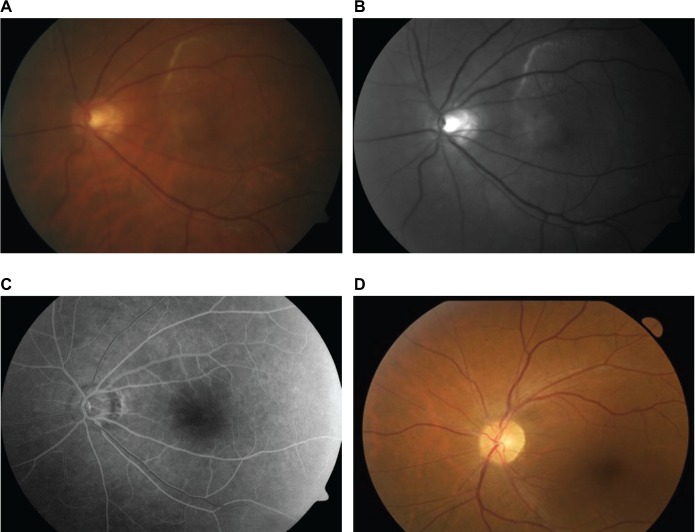
Figure 1.
Left fundus photographs of a 74-year-old male.
Notes: (A) Fundus at presentation showing a creamy-white lesion above the superior arcade and extending to involve the macular. (B) Red-free filter of the same image allowing visual enhancement of the lesion. (C) Venous phase angiogram identifying disk hyperfluorescence. (D) Fundus photograph of the same eye following treatment completion with IV benzylpenicillin.
Abbreviation: IV, intravenous.

