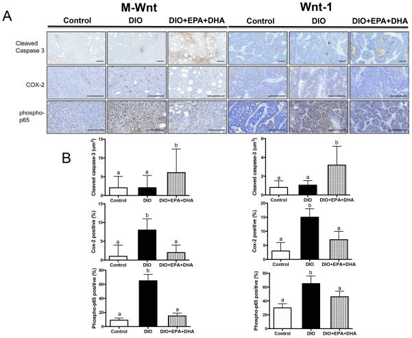
Figure 4.
The effect of a DIO regimen +/− EPA+DHA supplementation on IHC tumoral staining. A, representative photomicrographs of pathology and IHC staining of tumors for cleaved caspase-3, COX-2 and phospho-p65. B, bar graphs representing the Aperio image quantitation, scale bars indicate 100 μm, n=6 per group. Means ± SEM, statistically significant (P<0.05) differences are indicated by different letters.

