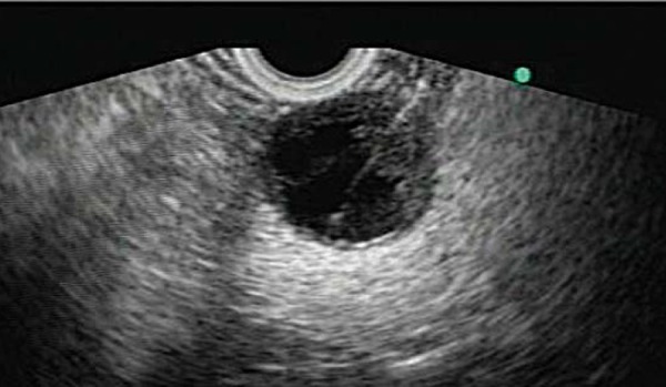
Fig. 10.

A neuroendocrine tumour in the body of the pancreas, FNA being performed. Note the sharp bordered, small, rounded configuration with a combination of cystic and solid elements – this is typical of the EUS appearance of neuroendocrine tumours.
