Fig. 14.

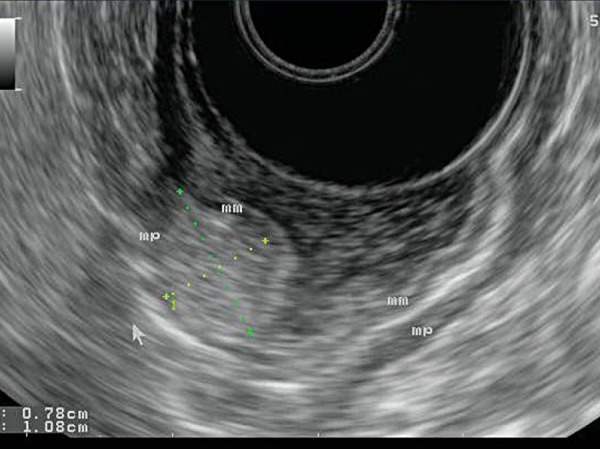
A lipoma arising from within the gastric wall. Endoscopically, this would look the same as the GIST seen in Fig. 13. However, EUS allows differentiation between the two: the lipoma is brightly hyperechoic.

A lipoma arising from within the gastric wall. Endoscopically, this would look the same as the GIST seen in Fig. 13. However, EUS allows differentiation between the two: the lipoma is brightly hyperechoic.