Figure 3b.

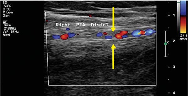
CDU image of an occluded posterior tibial artery with thrombus and serpiginous, collateral flow in and around the vessel. (“corkscrew appearance”). 5 Unlike vasculitis, the occluded vessel walls remain unchanged (indicated by arrows).

CDU image of an occluded posterior tibial artery with thrombus and serpiginous, collateral flow in and around the vessel. (“corkscrew appearance”). 5 Unlike vasculitis, the occluded vessel walls remain unchanged (indicated by arrows).