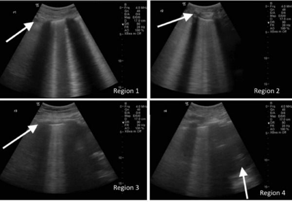
Figure 3.

A good quality scan set contained appropriate labels, a high focus and a low depth. A set order was required to mitigate poor labelling (R1 to 4 then L1 to 4) and when dubious this was crosschecked against subtle differences in the views, as indicated by block arrows. Region 1 usually contains either clavicle or subclavian vessel in the top left corner, region 2 has more rounded or cartilaginous rib, region 3 has fewer or less distinct rib shadows due to oblique rib position in axilla, and region 4 requires a moiety of either diaphragm, liver or spleen, to demonstrate that the sonologist has scanned the lowermost portion of lung posterolaterally. This scan set is strongly positive.
