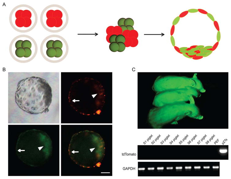
Figure 4.
Construction of chimeric porcine embryos by embryo aggregation. (A): A schematic diagram showing production of chimeric blastocysts using GFP/tdTomato-expressing four-cell stage embryos. (B): Both induced pluripotent stem cell (iPSC)-derived and porcine fetal fibroblast-derived blastomeres were incorporated into the inner cell mass (arrow head) as well as the trophectoderm (arrow) of blastocysts 4 or 5 days after aggregation. Scale bar =50 μm. (C): One-week-old piglets derived from aggregated embryos. Only green fluorescence could be detected in newborns, indicating that iPSC-derived cells did not contribute to late stage chimera formation. PCR analysis using pZT-specific primers (for iPSCs) did not detect any contribution of pig iPSCs in eight fetuses (S1–S8).

