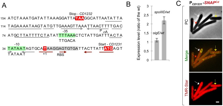
Fig 2. Constitutive expression of the skinCd gene CD1231.
A: promoter region of the CD1231 gene. The mapped transcriptional start sites (+1, red) [34] and the -10 and -35 promoter elements (green boxes) that match the consensus for σA recognition are indicated. Also represented are the stop codon of CD1232 and the start codon of CD1231 (red). Inverted repeats upstream (grey arrows) and downstream (brown) of the +1 position are indicated as well as a possible RBS overlapping the left arm of these repeats. B: qRT-PCR analysis of CD1231 transcription in strain 630Δerm, and in sigE or spoIIID mutant. RNA was extracted from cells collected 14 h (sigE mutant) or 15 h (spoIIID mutant) after inoculation in liquid SM. Expression is represented as the fold ratio between the indicated mutants and the wild-type (WT). Values are the average ± SD of two independent experiments. C: cells of the C. difficile 630Δerm strain carrying a PCD1231-SNAPCd transcriptional fusion were collected after 24 h of growth in liquid SM, stained with TMR-Star and the membrane dye MTG, and examined by phase contrast (PC) and fluorescence microscopy. The merged images show the overlap between the TMR-Star (red) and MTG (green) channels. The yellow arrow shows a vegetative cell expressing PCD1231-SNAPCd, the white arrow shows expression in the forespore and the red arrow expression in the mother cell. Scale bar, 1 μm.

