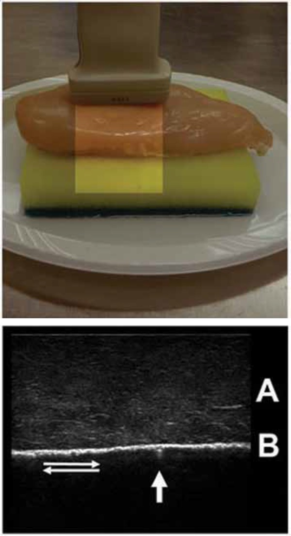
Figure 7.

A simple normal lung phantom and corresponding ultrasound image – chicken breast and synthetic sponge.
A: The simulated chest wall (chicken breast)
B: The pleural interface with opposing parietal and visceral pleura (synthetic sponge wrapped in plastic cling film to allow easy lung sliding) An assistant slides the sponge up and down the back of the simulated chest wall whilst the trainee scans. This simulates the normal ventilating lung. Sliding occurs between the two pleural surfaces as the diaphragm descends and ascends with each respiratory cycle.
Note the small comet tail artifact (arrow). In normal lung these are few and weak fading within a few millimetres of the pleural surface. They probably represent reverberation of ultrasound within tiny pockets of interstitial fluid. Watch also for lung sliding with each respiratory cycle (double arrows).
Numerous pathologies including non‐ventilating lung, bullae and pleuradhesis may disrupt the normal appearance.
