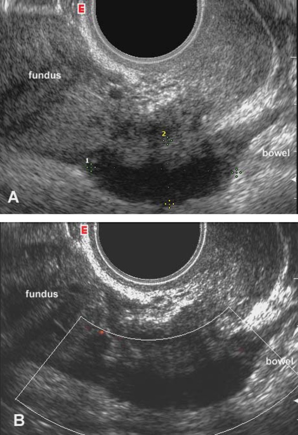
Figure 1.

Transvaginal sagittal scan of the posterior compartment of the pelvis, including the pouch of Douglas. A) Greyscale image: a solid hypoechoic nodule with blurred margins and a hyperechoic rim (calipers) suggestive of the presence of DIE is seen at the level of the anterior wall of the rectum. B) Power Doppler reveals the absence of blood vessels inside the implant.
