
Figure 3.
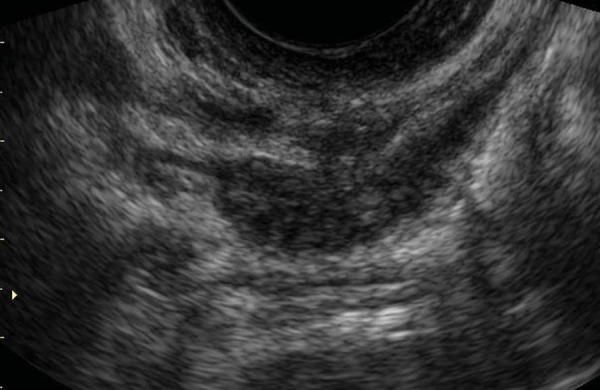
Transvaginal sagittal scan of the posterior compartment of the pelvis, showing the presence of an endometriotic nodule extending caudally toward the rectovaginal septum. DIE appears as a solid hypoechoic nodule with spiculated margins and a hyperechoic rim, close to the tip of the vaginal probe.