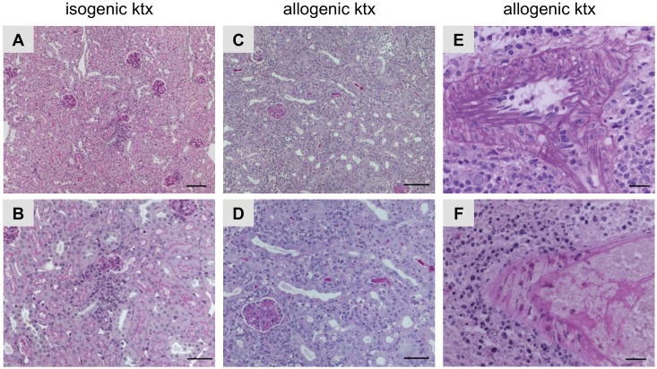
Fig 1. Histopathology in isogenic and allogenic kidney grafts.
PAS stainings illustrating histopathology of mouse isografts (A, B) and allografts (C-F). Isografts showed almost normal renal morphology with some focal interstitial inflammatory infiltrates (A and B). Mouse allografts revealed severe rejection (C and D). In addition, allografts with arterial endothelialitis (Banff IIA, E) and transmural arteriitis (Banff III, F) are shown. Bars represent 20 μm in A and B, 50 μm in B and D and 100 μm in E and F.

