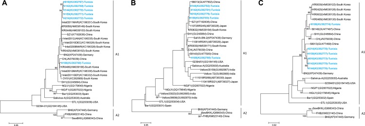
Fig 3. Phylogenetic trees of saliviruses detected in diarrheal Tunisian children.
A. VP0 region of salivirus (815 nt); B. 2CHel region of salivirus (275 nt); C. 3Dpol region of (686 nt); Phylogenetic trees were inferred using the Maximum Likelihood method based on the Tamura-3-parameter nucleotide substitution model with a discrete gamma distribution. Bootstraps values were calculated from 1000 replicates. Strains of this study are shown in blue. Genotypes are shown in bold.

