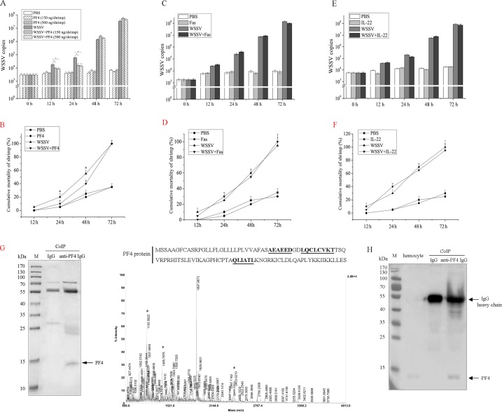
Fig 2. The roles of cytokines in the antiviral immunity of shrimp.
(A) The influence of PF4 on WSSV copies in shrimp. The shrimp were simultaneously injected with WSSV and PF4 (at various concentrations). At different time points post-infection, the shrimp were subjected to quantitative real-time PCR to quantify the WSSV copies. As controls, PBS, PF4 alone (at various concentrations) and WSSV alone were included in the injections. (B) The effects of PF4 on the mortality of WSSV-infected shrimp. The shrimp were simultaneously injected with WSSV and PF4 (150 ng/shrimp). PBS, PF4 alone (at various concentrations) and WSSV alone were included in the injections as controls. At different times after injection, the cumulative mortality of shrimp was monitored. (C) The role of Fas in the virus infection of shrimp. The shrimp were simultaneously injected with WSSV and Fas (150 ng/shrimp). At different times post-infection, the WSSV copies of shrimp were quantified by quantitative real-time PCR. As controls, PBS, Fas alone and WSSV alone were included in the injections. The numbers indicated the time points post-infection. (D) The influence of Fas on the WSSV-infected shrimp mortality. At different times after injection of WSSV+Fas, the cumulative mortality of shrimp was examined. PBS, Fas alone and WSSV alone were used as controls. (E) The impact of IL-22 on WSSV copies in shrimp. The shrimp were treated with WSSV+IL-22 (150 ng/shrimp), PBS, IL-22 alone or WSSV alone, followed by the detection of WSSV copies using quantitative real-time PCR. The numbers indicated the time points post-infection. (F) The effects of IL-22 on the mortality of WSSV-infected shrimp. The cumulative mortality of shrimp treated with WSSV+IL-22 (150 ng/shrimp), PBS, IL-22 alone or WSSV alone was evaluated at different times post-infection. (G) The identification of PF4 protein in shrimp. Coimmunoprecipitation (CoIP) assays were conducted using shrimp hemocytes with anti-PF4 IgG (left panel). Rabbit IgG was used as a control. The proteins were analyzed using SDS-PAGE with Coomassie brilliant blue staining. Then the protein was identified by mass spectrometry (right panel). The matched peptides were underlined. M, protein marker. (H) Western blot detection of PF4 protein in the shrimp hemocytes and in the CoIP products. The proteins in the shrimp hemocytes and in the CoIP products were separated by SDS-PAGE. Then Western blotting was conducted using the antibody against the PF4 protein. The proteins were indicated by arrows. M, protein marker. In all panels, statistically significant differences between treatments are represented with asterisks (*, p<0.05).

