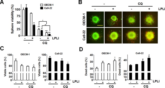
Fig 6. LPLI reduced tumor viability in spheroid culture.
(A) OECM-1 or Ca9-22 cells were sphere cultured and then exposed to LPLI (810 nm, 60 J/cm2) in the presence or absence of CQ (20 μM) for 48 h. The spheres were lysed to measure ATP level for cell viability. (B) The viable and dead spheres as cultured and treated as (A) were imaged with LIVE (green)/DEAD (red) staining kit. Representative data are shown. Scale bar: 400 μm. (C) The green and (D) red fluorescence of the spheres as (B) was quantitated with a reader for the viable and dead cell population, respectively (n = 6). The quantified results are expressed as the mean ± SEM from 3 individual experiments. n.s., p > 0.05; *p < 0.05; **p < 0.01; ***p < 0.001.

