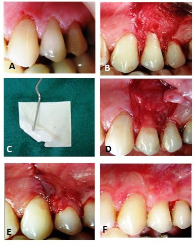
Figure 4.

Test group: A) Recession defect on the vleft maxillary bicuspid; B) Preparation of the recipient site; C) Preparation of the amniotic membrane; D) The amniotic membrane was placed over the exposed root surface; E) The flap was positioned coronally over the amniotic membrane and secured with a sling sutures; F) Partial root coverage at 6-month follow-up.
