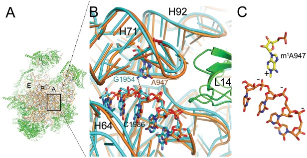
Fig 4. High structural conservation between S. scrofa mitoribosome and E. coli ribosome at position 947.
(A) The structure of the porcine mitoribosomal large subunit (PDB accession code 4v1a and 4v19) shown from the subunit interface side. The ribosomal RNA is shown in brown and the ribosomal proteins in green. The ribosomal tRNA A-, P-, and E-binding sites are indicated. (B) Sticks-and-ribbon representation of interaction between helices H71 and H64 in S. scrofa (brown) mitoribosome or E. coli (turquoise) ribosome (PDB accession code 4ybb). The hydrogen bond that is likely disrupted by an adenine in position 947 is represented as a dashed line. (C) The positively charged m1A947 stabilizes the structure by interacting with the negatively charged H64 backbone. Numbers refer to the positions of E. coli ribosomal RNA.

