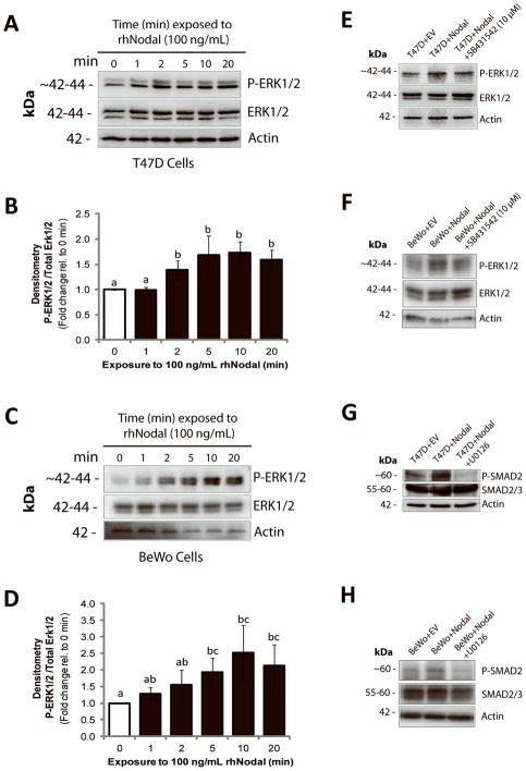
Figure 3. Nodal activates ERK signaling.
(A) Western blot time course analysis of ERK1/2 activation in T47D cells following treatment with 100 ng/mL rhNodal for 0, 1, 2, 5, 10, or 20 minutes. P-ERK1/2 increases compared to controls after 2 minutes of rhNodal treatment. Total ERK1/2 and β-Actin are used as controls. (B) Densitometric analysis for all replicate experiments corresponding to (A). ImageJ was used to calculate band density of P-ERK1/2 relative to total ERK1/2. Data are presented as mean fold change ± S.E.M. Different letters indicate a significant difference compared to controls (n=4, p=0.029). (C) Western blot time course analysis of ERK1/2 activation in BeWo cells following treatment with 100 ng/mL rhNodal for 0, 1, 2, 5, 10, or 20 minutes. P-ERK1/2 increases compared to controls after 5 minutes of rhNodal treatment. Total ERK1/2 and β-Actin are used as controls. (D) Densitometric analysis for all replicate experiments corresponding to (C). ImageJ was used to calculate band density of P-ERK1/2 relative to total ERK1/2. Data are presented as mean fold change ± S.E.M. Different letters indicate a significant difference compared to controls (n=4, p=0.029). (E) Western blot demonstrating that P-ERK1/2 is elevated in T47D cells transfected with a Nodal expression vector (T47D+Nodal) versus a control vector (T47D+EV), and that phosphorylation is reduced when T47D+Nodal cells are treated with SB431542 (10 μM). Total ERK1/2 and β-Actin are used as controls. (F) Western blot demonstrating that P-ERK1/2 is elevated in BeWo cells transfected with a Nodal expression vector (BeWo+Nodal) versus a control vector (BeWo+EV), and that phosphorylation is reduced when BeWo+Nodal cells are treated with SB431542 (10 μM). Total ERK1/2 and β-Actin are used as controls. (G) Western blot demonstrating that P-SMAD2 is elevated in T47D+Nodal cells compared to T47D+EV cells, and that this effect is abrogated by treating T47D+Nodal cells with 10 μM U0126 (1 hr). Total SMAD2/3 and β-Actin are used as controls. (H) Western blot demonstrating that P-SMAD2 is elevated in BeWo+Nodal cells compared to BeWo+EV cells, and that this effect is abrogated by treating BeWo+Nodal cells with 10 μM U0126 (1 hr). Total SMAD2/3 and β-Actin are used as controls.

