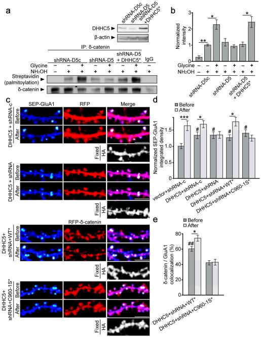
Figure 8. DHHC5 is required for activity-induced palmitoylation of δ-catenin and surface AMPAR insertion.
(a top) shRNA-D5 reduced DHHC5 levels at 12 DIV (shRNA-D5c[1.0 ± 0.13]: shRNA-D5 [0.62 ± 0.042], shRNA-D5+DHHC5* [0.89 ± 0.044]; one-way ANOVA, p=0.042, F2,6=5.57). (a bottom, b) Knockdown of DHHC5 abolishes activity-induced palmitoylation of δ-catenin (n=3 blots from 3 separate cultures, p=0.002, F6,14=10.03). (c) Confocal images of hippocampal neurons transfected with the indicated constructs before and 40–60 min after glycine treatment. SEP-fluorescent puncta are pseudocolored in heat maps. DHHC5-HA overexpression was confirmed by post hoc immunostaining for HA. Scale bar=5μm. (d) IntDen of SEP-GluA1 puncta, normalized to the same puncta before glycine treatment. n values indicate cell number from ≥3 separate cultures, and p values from paired t-tests: vector+shRNA-c (14, p<0.001), DHHC5+δ-catenin shRNA-c (15, p=0.006), DHHC5+δ-catenin shRNA (13, p=0.857), DHHC5+δ-catenin shRNA+δ-catenin WT* (16, p=0.002), and DHHC5+δ-catenin shRNA+δ-catenin C960-1S* (13, p=0.161). DHHC5 overexpression increased basal SEP-GluA1 IntDen (one-way ANOVA; p=0.001, F9,132=4.141). Crosshatches denote significance among “before” groups relative to vector+shRNA-c, asterisks denote significance within groups before and after glycine. (e) Colocalization of indicated δ-catenin constructs with GluA1 (one-way ANOVA; p<0.001, F3,54=19.29). Crosshatches denote significance among “before” groups relative to DHHC5+shRNA+C960-1S*. WT*/GluA1 colocalization increased following activity (paired t-test; p=0.005), whereas C960-1S*/GluA1 colocalization did not (paired t-test; p=0.741). Graphs represent mean ± SEM. (b) *p<0.05, one-way ANOVA with Tukey’s test post hoc. (d, e) *p<0.05, ***p<0.001, paired t-test; #p<0.05, ##p<0.01, one-way ANOVA with Tukey’s test post hoc. Full-length blots from (a) are presented in Supplementary Figure 6.

