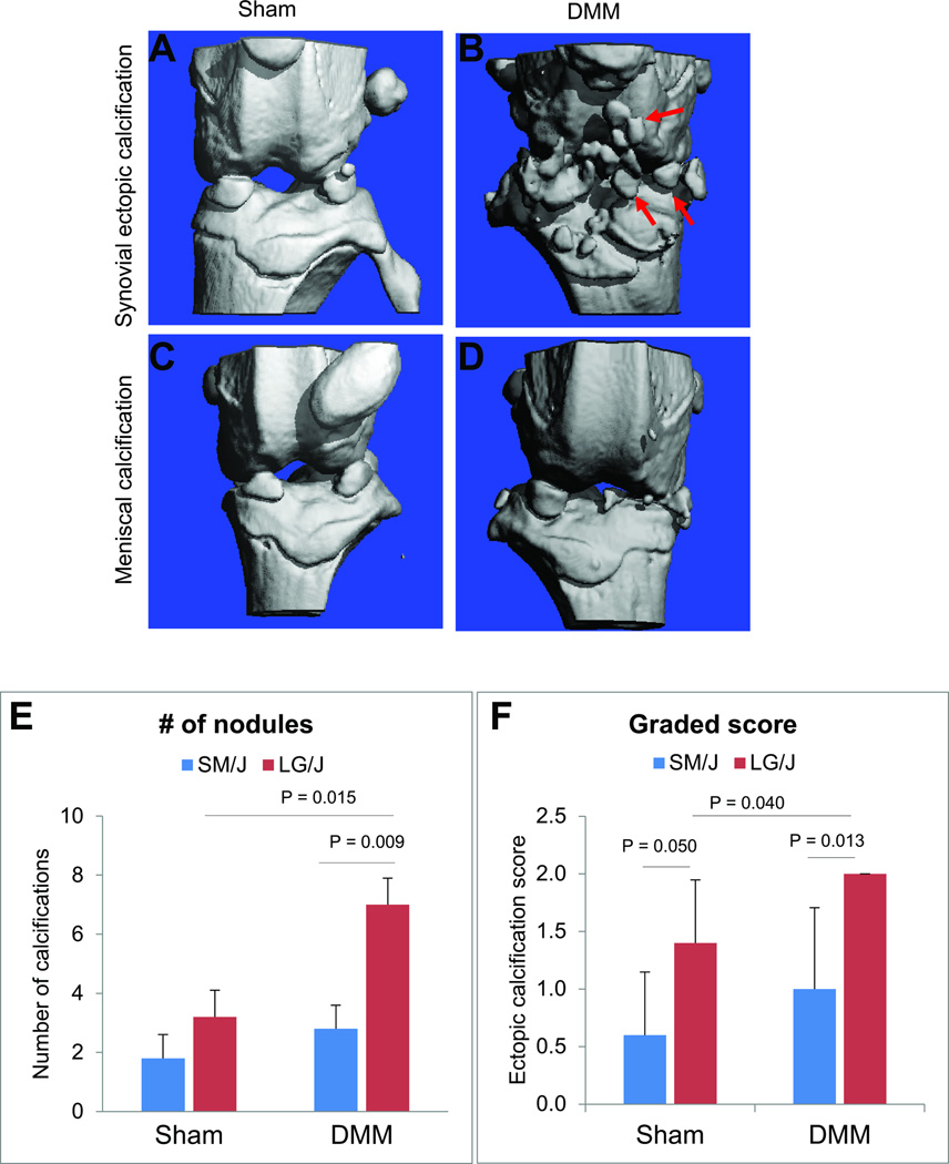
Fig. 2. Typical phenotypic outcomes in sham and DMM knees.
F44 advanced intercross line mice were subjected to DMM at 10-week of age. Micro-CT analysis on the harvested knees was performed 8-week post-surgery. Micro-CT images showed ectopic synovial (A–B) and meniscal (C–D) calcifications exclusively in the knees subjected to DMM. No significant calcifications were observed in contralateral sham knees (A, C). We observed that ectopic synovial calcifications were significantly higher in the knees subjected to DMM of LG/J compared to contralateral sham-operated knee as well as DMM-operated knee of SM/J strain (E–F).

