


Figure 2.
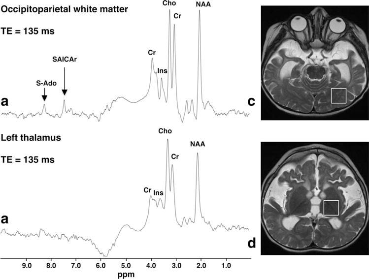
Single-voxel proton MRS (TR/TE 1500/135 msec) obtained at 1.5 T from the left occipitoparietal region (a) and thalamus (b) of patient 1 at the age of 18 months. Corresponding ROIs are shown in (c,d), respectively. The thalamus spectrum (b) is unremarkable, but the WM spectrum (a) clearly shows abnormally elevated peaks at 8.3 and 7.5 ppm, which are assigned to S-Ado and SAICAr, respectively.