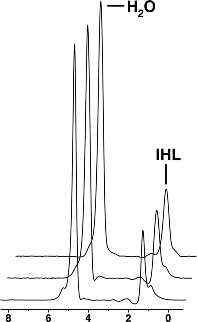
Figure 1.
1H-MRS of the right hepatic lobe in a 36 year-old man obtained at baseline (A), 6 weeks (B), and 6 months (C). Intrahepatic lipid (IHL) content by 1H-MR spectroscopy shows low variation between scans (fat fraction 23.7% at baseline, 24.3% at 6 weeks and 24.7% at 6 months). For purposes of visual comparison, the amplitudes of unsuppressed water are scaled identically.

