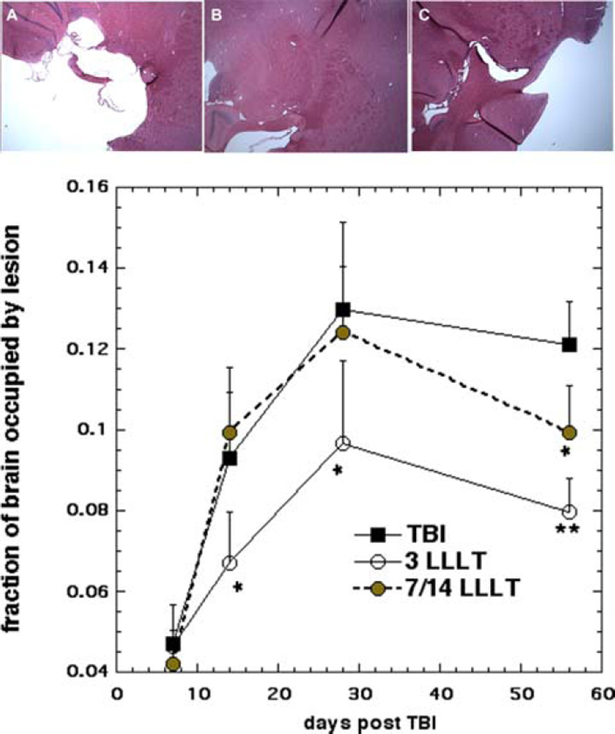
Figure 3.
(A–C) Sample H&E stained sections from mouse brains. Mice were sacrificed at 56 days after having received; (A) zero laser treatments, TBI; (B) 3 laser treatments; or (C) 14 laser treatments. (D) Lesion size measurement. Mice (n = 4) were sacrificed at 1 week, 2 weeks, 4 weeks and 8 weeks, and lesion size determined from H&E stained sections. Data points are mean ± SD (n = 4) of the fraction of total brain occupied by lesion. *p < 0.05 vs. untreated TBI and 14 LLLT; *p < 0.05 vs. untreated TBI; **p < 0.01 vs untreated TBI.

