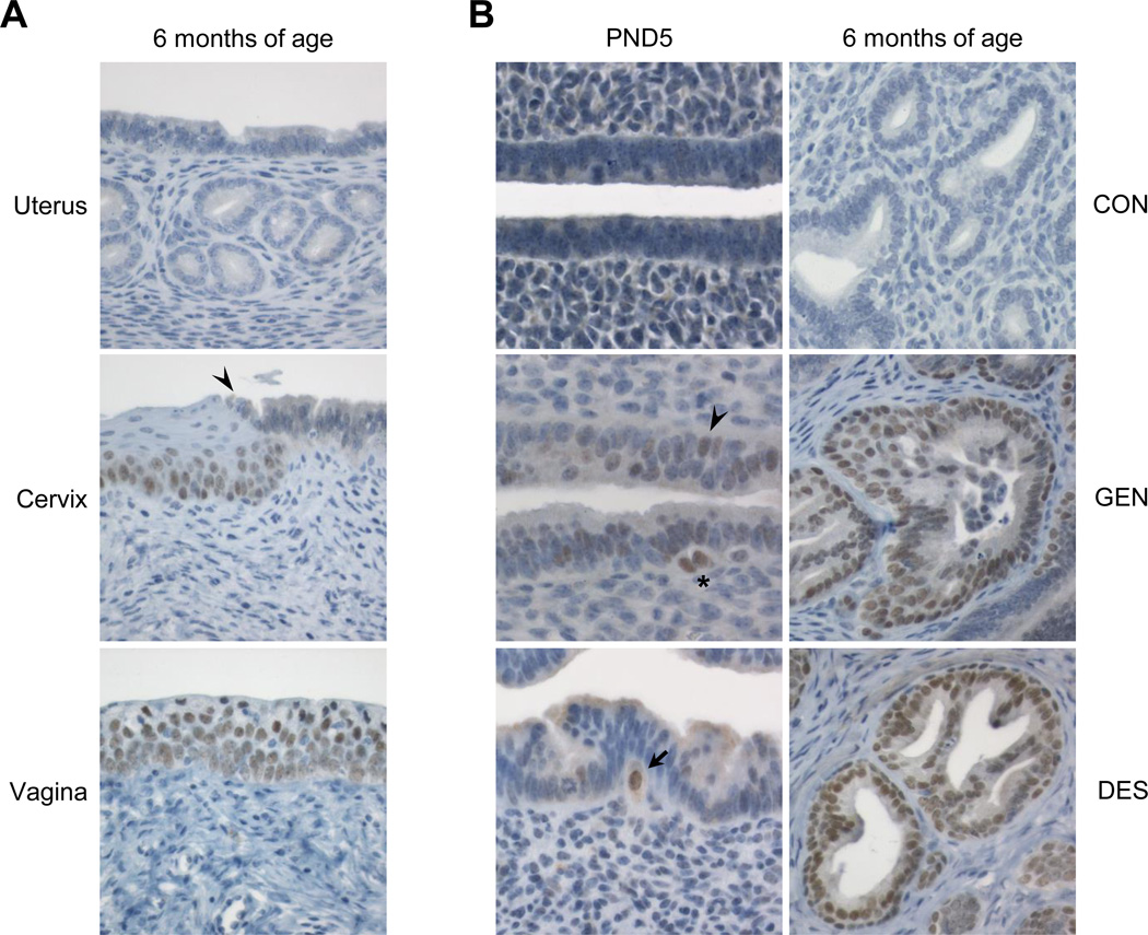
Figure 1.
SIX1 localization in controls and following neonatal estrogenic chemical exposure. A. Representative SIX1 immunolabeling in a control adult female mouse reproductive tract at 6 months of age. Arrowhead indicates squamocolumnar junction (SCJ). B. Appearance and expansion of SIX1 immunolabeled cells in mouse endometrium following neonatal GEN or DES exposure at PND5 or 6 months of age. Arrowhead indicates SIX1-positive columnar cells and asterisk indicates SIX1-positive basal-type cells underlying the glandular epithelium. Arrow indicates large SIX1-positive basal-type cell that appears to be traversing the basement membrane. Representative images were taken at an objective magnification of 60× (PND 5) or 40× (6 months of age).

