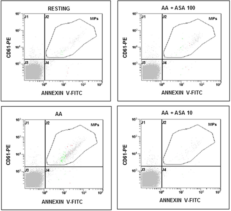
FIGURE 1.

Flow cytometric analysis of platelet-derived microparticles shed after platelet stimulation with arachidonic acid (AA; 1.25 mmol/L) and in the presence of different concentrations of ASA (100 and 10 μmol/L). Dot plots show the dual fluorescence analysis of representative PFP stained with Annexin V-fluorescein isothiocyanate (FITC) and anti-CD61-phycoerythrin (PE). The total number of CD61+ MPs was calculated as the sum of CD61+/Annexin and CD61+/Annexin + MPs. Absolute counts of PMPs were determined by using Flow-CountTM Fluorospheres (Beckman Coulter) and expressed per microliter of PFP. Experiments were performed in duplicate.
