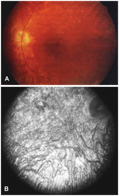
Figure 1.

Fundoscopy of JNCL patient eyes. (A) Posterior pole of the left eye of an 8-year-old female JNCL patient with the common 1 kb deletion showing bull’s eye maculopathy, attenuated vessels, and pale optic discs. Reproduced, with permission, from Ref. 50. (B) Angiogram with diffuse stippled hyperfluorescence in the eye of a 9-year-old JNCL patient. Reproduced, with permission, from Ref. 47.
