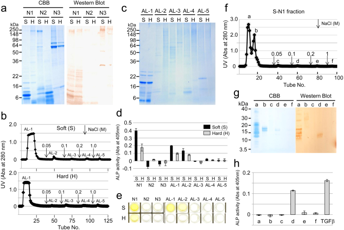
Figure 2. TGF-β1 in porcine enamel matrix.
(a) SDS-PAGE (15% gel) stained with Simply Blue (CBB) (left) and western blot (right) using a specific antibody against amelogenin, showing the neutral soluble extracts from soft (S) and hard (H) enamel fractionated by successive ammonium sulfate precipitations. N1: ammonium sulfate precipitate (ASP) at 40% saturation, N2: 40–65% ASP soluble in acid and N3: 40–65% ASP insoluble in acid. (b,f) Heparin Sepharose chromatograms showing the absorbance at 280 nm for (b) AL extracts from soft enamel (~200 mg) (Soft) and hard enamel (~150 mg) (Hard), and (f) S-N1 extracts (~150 mg) from soft enamel. Downward-pointing arrows represent the starting points of the step gradient with 0.05, 0.1, 0.2 and 1 M NaCl. (c) SDS-PAGE (15% gel) stained with Simply Blue (CBB) showing fractions AL-1 to AL-5 on heparin chromatograms from soft (S) and hard (H) enamel. (d,h) ALP-inducing activity of HPDL cells exposed to (d) fractions N1 to N3 and AL-1 to AL-5 from soft (S) and hard (H) enamel, and (h) fractions a-f isolated from heparin chromatography. The recombinant human TGF-β1 with a carrier (0.3 ng mL−1) (TGFβ) was used as a positive control for the detection of ALP-inducing activity of HPDL cells (n = 9 culture wells for each sample). (e) ELISA for the detection of TGF-β1 capture and TGF-β1 detection antibodies in fractions N1 to N3 and AL-1 to AL-5 from soft (S) and hard (H) enamel. (g) SDS-PAGE (15% gel) stained with Simply Blue (CBB) (left) and western blot (right) using a specific antibody against amelogenin, showing fractions a-f isolated from heparin chromatography.

