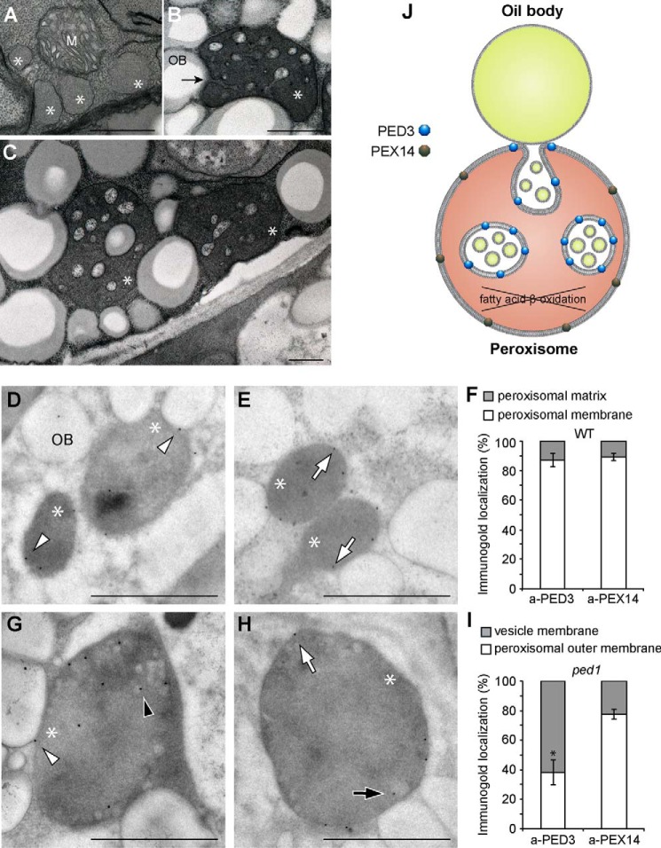
FIGURE 7.
Membrane localization of PED3 in the wild type and the ped1 mutant. A–C, peroxisomes in the etiolated cotyledons of 5-day-old wild-type (A) and ped1 mutant (B and C) seedlings. The arrow indicates membrane invagination of the enlarged peroxisomes at the interaction site with an oil body. Scale bars = 0.5 μm. M and OB indicate mitochondrion and oil body, respectively. Asterisks indicate the peroxisomes. D, E, G, and H, immunogold labeling of ultrathin sections of etiolated cotyledons from the wild type (D and E) and the ped1 mutant (G and H). Immunogold particles were coated with antibodies raised against PED3 (D and G) and PEX14 (E and H). Scale bars = 0.5 μm. Arrowheads and arrows indicate PED3- and PEX14-coated gold particles, respectively, on peroxisomal outer membranes (white) and vesicle membranes (black). F and I, quantitative analysis of the membrane localization of PED3 and PEX14 in the wild type (F) and ped1 mutant (I) based on immunogold labeling. The y axis represents the percentage of immunogold localization on different parts of peroxisomes. Data are derived from supplemental Table 1 and represent the mean ± S.D. of 10 sections (see also supplemental Table 1). * indicates a statistically significant difference between PED3 and PEX14 localization (p < 0.01, Student's t test). J, schematic showing the enrichment of PED3 and PEX14 on different membranes of enlarged peroxisomes in ped1.

