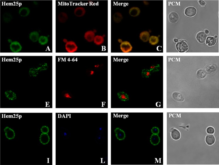
FIGURE 3.
Subcellular localization of recombinant Hem25p after expression in S. cerevisiae cells. hem25Δ cells overexpressing the Hem25p/V5 protein were grown as described under “Experimental Procedures.” Staining of the Hem25p/V5 protein was performed with mouse anti-V5 monoclonal antibody and anti-mouse antibody with conjugated FITC (A, E, and I). MitoTracker Red (B), FM 4-64 (F), and DAPI (L) were used to locate mitochondria, vacuoles, and nucleic acid in the cells, respectively. Colocalization of the Hem25p/V5 protein and mitochondria are seen as yellow fluorescence in the red and green merged image (C). Phase contrast microscopy (PCM) was used to monitor the integrity of the cells (D, H, and N). The same cells were photographed first with a FITC-green filter set and then with the MitoTracker or FM 4-64 red filter set or with the DAPI blue filter set. Identical fields are presented in panels A--D, E–H, or I–N.

