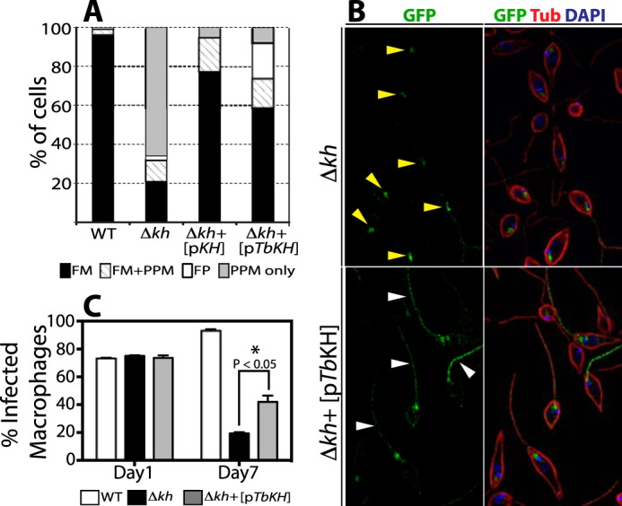
FIGURE 11.

Functional complementation of Leishmania kharon null mutant by TbKH. L. mexicana Δlmxkh null mutants (Δkh) expressing the LmxGT1 flagellar glucose transporter tagged with GFP at it C terminus [pGT1::GFP] were transfected with an episomal expression vector encoding the TbKH gene [pTbKH]. Δlmxkh[pGT1::GFP] (Δkh) and Δlmxkh[pGT1::GFP]/pTbKH (Δkh+[pTbKH]) were stained with DAPI (blue) and immunostained with anti-GFP pAb (GFP, green) and anti-α-tubulin mAb (Tub, red). A, quantification of LmxGT1::GFP localization (%) in L. mexicana lines. The data for WT L. mexicana, Δkh, and Δkh[pKh] (Δkh null mutants complimented with an episomal expression vector encompassing the LmxKH gene) were previously published by Tran et al. (21) and are reproduced here for comparison. The key at the bottom shows the labeling scheme for quantification of parasites that contain GT1::GFP in the flagellar membrane (FM), flagellar membrane plus pellicular plasma membrane (FM+PPM), flagellar pocket (FP), and pellicular plasma membrane only (PPM only). The number of parasites examined in each quantification was: WT = 210, Δkh = 241, Δkh+[pKH] = 139, and Δkh+[pTbKH] = 249. B, examples of LmxGT1::GFP localization in non-complemented (Δkh, upper panel) and complemented (Δkh+[pTbKH]) mutants. Yellow arrowheads in the upper panel indicate GT1::GFP localized in the flagellar pocket. White arrowheads in the lower panel indicate GT1::GFP in the flagellum. C, quantification of THP-1 infection with WT, Δkh mutant, and Δkh complemented with an episomal copy of TbKH Δkh+[pTbKH] promastigotes. Day 1 and Day 7 indicate the time elapsed following infection. The data represent the averages and standard deviations of triplicate infections (n = 3), each counted in triplicate and averaged. The statistical significance for comparison of the null mutant and complemented null mutant on day 7 was determined by two-tailed Student's t test (p < 0.05).
