
FIGURE 9.
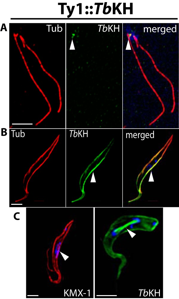
s Subcellular localization of TbKH in PF parasites. A, flagellar axonemes were isolated from Ty1::TbKH-expressing PF parasites by detergent lysis and incubation with NaCl (“Experimental Procedures”) to disrupt the subpellicular microtubules, and preparations were stained with antibodies against α-tubulin (Tub, red) and Ty1 (BB2 mAb, green) and with DAPI to visualize kDNA (blue). The white arrowhead indicates the green fluorescence representing Ty1::TbKH at the base of the flagella. B, Ty1::TbKH PF parasites were immunostained with BB2 mAb (TbKH, green) and anti-α-tubulin (Tub, red) to show localization of TbKH that partially overlaps with the subpellicular microtubules (white arrowhead). C, Ty1::TbKH PF parasites were stained with DAPI (blue) and immunostained with BB2 mAb (TbKH, green) or KMX-1 mAb (Tub, red). The white arrowhead indicates KMX-1 antibody staining on the spindle between two nuclei (left image) and TbKH on the mitotic spindle (right image) in parasites undergoing karyokinesis.