
FIGURE 6.
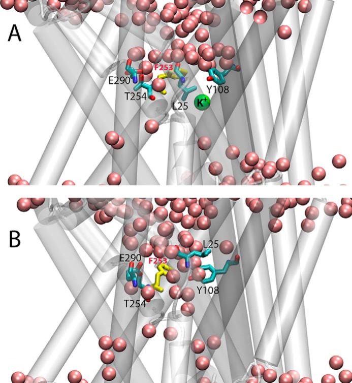
Snapshots from initial (A) and final (B) frames of OCCnK trajectory illustrating different orientation of Phe253 residue (in yellow) and formation of continuous water channel (pink spheres) connecting EC vestibule to the functional sites of LeuT. Glu290, Thr254, Leu25, and Tyr108 residues are shown in licorice rendering, and K+ ion is depicted as a green sphere.