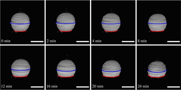
FIGURE 7.
Flattening of a GUV with elevated membrane tension after ENTH binding. Time series of three-dimensional reconstructions of SDCLM images of a GUV (DOPC/DOPE/cap-biotin-PE/Atto488-DPPE/PtdIns(4,5)P2, 66.2:30:2:1:0.8) adhering on an avidin-coated glass surface generating high membrane tension (c(Mg2+) = 2 mm). The GUV starts to flatten after ENTH addition (cENTH = 1 μm), and Ri/Rad increases. Flattening starts at t = 0 min. Scale bars: 20 μm.

