FIGURE 3.
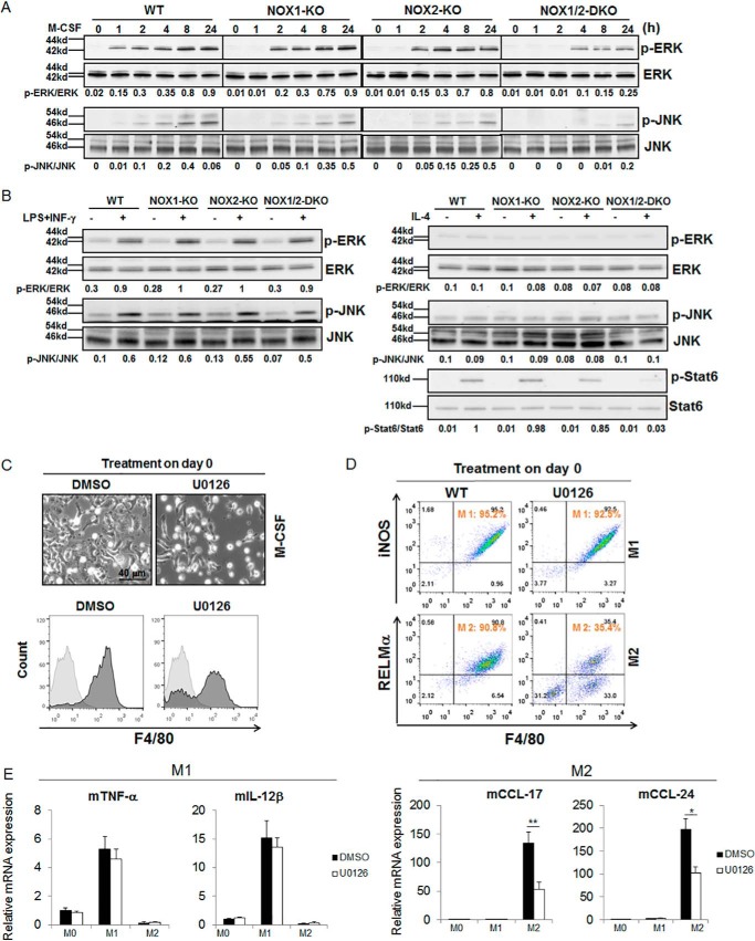
NOX1 and NOX2 depletion inhibited ROS-mediated ERK and JNK activation. A, mouse BMMs from WT, NOX1-KO, NOX2-KO, and NOX1/2-DKO mice were treated with M-CSF (20 ng/ml) for the indicated time points. The expression levels of p-ERK, ERK, p-JNK, and JNK were determined by Western blotting. B, mouse monocytes from WT, NOX1-KO, NOX2-KO, and NOX1/2-DKO mice were treated with M-CSF for 6 days. On day 6, cells were treated with LPS/INF-γ (left panel) or IL-4 (right panel) for 15 min. Cell lysates were immunoblotted with the indicated antibodies. C, on day 0, mouse monocytes from WT, NOX1-KO, NOX2-KO, and NOX1/2-DKO mice were treated with DMSO or ERK inhibitor (U0126, 5 μm) for 1 h and then treated with M-SCF for 6 days. On day 6, the cells were analyzed by imaging and flow cytometry. Representative light microscopy image are shown (top panel). Macrophages were collected and stained with anti-F4/80 antibody and analyzed by flow cytometry (bottom panel). D and E, on day 6 the macrophages from C were further treated for 24 h with either LPS (100 ng/ml) and INF-γ (20 ng/ml) for M1 or IL-4 (20 ng/ml) for M2 polarization. The M1 cell population (iNOS and F4/80) and M2 cell population (RELMα and F4/80) were detected by flow cytometry (D). Detection of M1 cytokines (TNF-α, IL-12β) or M2 chemokines (CCL17, CCL24) was by real-time PCR (E). *, p < 0.05; **, p < 0.001 versus WT.

