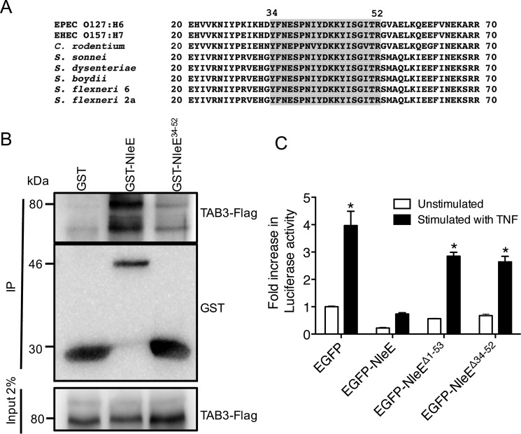
FIGURE 2.
Functional analysis of the TAB3-binding domain of NleE. A, alignment of the N-terminal regions of NleE and OspZ from A/E pathogens and Shigella. Amino acids 34–52 are shaded. B, pulldown of TAB3-FLAG by immobilized GST, GST-NleE, and GST-NleE34–52. C, -fold increase in NF-κB-dependent luciferase activity in HeLa cells expressing EGFP, EGFP-NleE, EGFP-NleEΔ1–53, or EGFP-NleEΔ34–52 and left unstimulated or stimulated with TNF for 8 h where indicated. Results are the mean ± S.E. (error bars) of three independent experiments carried out in duplicate. *, significantly different from unstimulated HeLa cells expressing EGFP only (p < 0.0001, unpaired two-tailed t test). EHEC, enterohemorrhagic E. coli; IP, immunoprecipitation.

