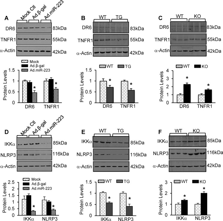
FIGURE 10.
A–C, representative Western blots and quantitative results of DR6 and TNFR1 protein expression in Ad.miR-223-infected rat cardiomyocytes (A), pre-miR-223 TG hearts (B), and pre-miR-223-null hearts (C) and their respective controls (n = 3 per group; *, p < 0.05 versus controls). D–F, protein levels of IKKα and NLRP3 were significantly reduced in Ad.miR-223-infected rat myocytes and pre-miR-223 TG hearts, whereas they were increased in pre-miR-223 KO hearts compared with the respective controls (n = 3 per group; *, p < 0.05 versus controls). Error bars represent mean ± S.D.

