
FIGURE 4.
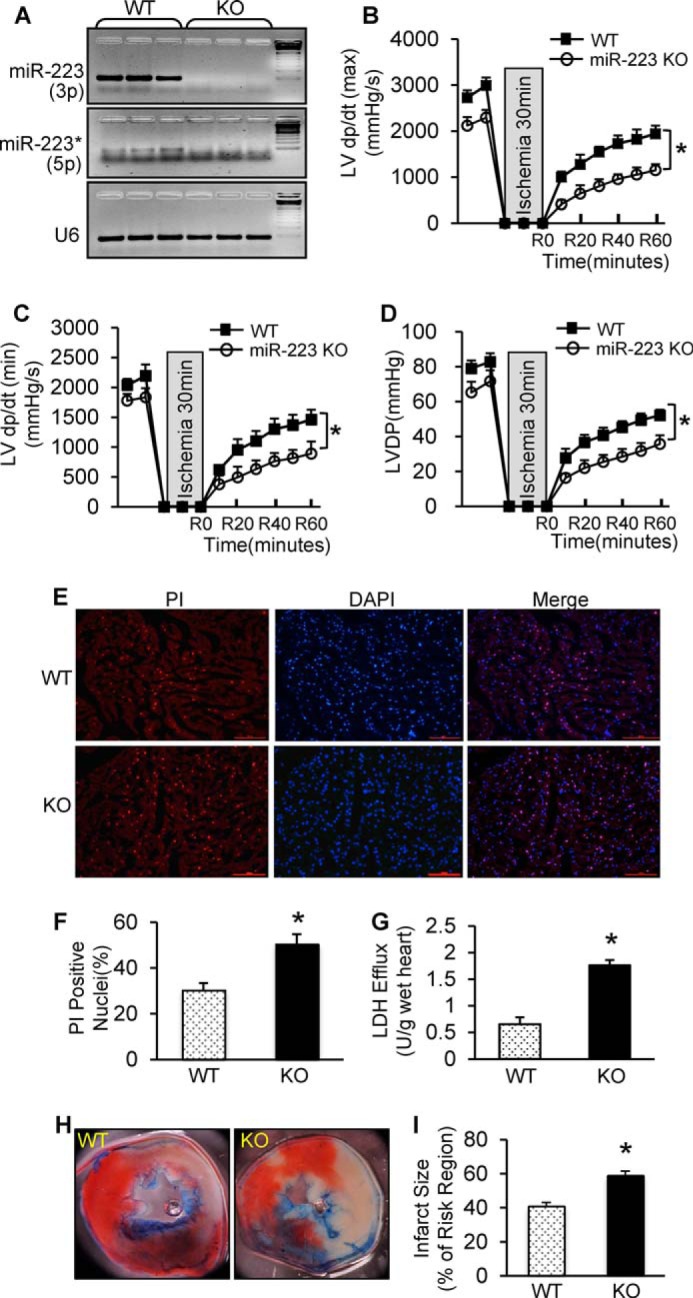
Ablation of pre-miR-223 aggravated ex vivo and in vivo I/R-induced cardiac injury. A, deletion of miR-223-3p/-5p expression was detected by semiquantitative RT-PCR. U6 was used as an internal control (n = 3 for each group). B–D, pre-miR-223 KO mice showed worse functional recovery than WTs during 1-h reperfusion. We performed 30-min ischemia for pre-miR-223 KO mice because KO mice could not bear 45-min ischemia (n = 8 for WT group; n = 9 for KO group; *, p < 0.005 versus WTs). E and F, pre-miR-223 KO hearts displayed a greater degree of myocardial necrosis than WTs as determined by PI staining (E) and quantitative analysis (F) (n = 8 for WT group; n = 9 for KO group; *, p < 0.05 versus WTs). G, total LDH in coronary effluent collected during the first 10 min of reperfusion was significantly increased in KO hearts compared with WTs (n = 8 for WT group; n = 9 for KO group; *, p < 0.05 versus WTs). H and I, pre-miR-223-null hearts exhibited a larger infarction size after 30 min of left anterior descending artery occlusion followed by 24 h of reperfusion. (n = 9 for WT group; n = 8 for KO group; *, p < 0.01 versus WTs). Error bars represent mean ± S.D.