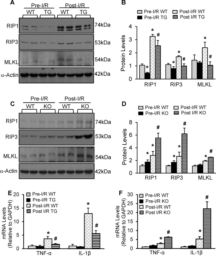
FIGURE 5.
MiR-223-3p/-5p duplex negatively regulated the RIP1/RIP3/MLKL necroptotic signaling cascades and the expression of inflammatory cytokines in I/R hearts. A and B, representative Western blots (A) and quantitative results (B) of RIP1, RIP3, and MLKL protein expression in WT hearts and pre-miR-223 TG hearts under in vivo pre-I/R and post-I/R (30 min/24 h) conditions (n = 6 hearts per experimental group; *, p < 0.05 versus pre-I/R WTs; #, p < 0.05 versus post-I/R WTs). C and D, representative Western blots (C) and quantitative results (D) of RIP1, RIP3, and MLKL protein expression in WT hearts and pre-miR-223 KO hearts under in vivo pre-I/R and post-I/R (30 min/24 h) conditions (n = 6 hearts per experimental group; *, p < 0.05 versus pre-I/R WTs; #, p < 0.05 versus post-I/R WTs). (Note: for the RIP1 immunoblotting, we initially used a rabbit anti-RIP1 antibody from Santa Cruz Biotechnology (in A), and the upper band was the real band of RIP1 (74 kDa); later on, we tried a rabbit anti-RIP1 from Cell Signaling Technology (in C)). E and F, the expression levels of IL-1β and TNF-α were analyzed by qRT-PCR in pre-miR-223 TG mouse hearts and WTs (E) and pre-miR-223 KO mouse hearts and WTs (F) subjected in vivo to 30-min ischemia followed by 4-h reperfusion. GAPDH mRNA was used as an internal control (n = 6; *, p < 0.05 versus pre-I/R WTs; #, p < 0.05 versus post-I/R WTs). Error bars represent mean ± S.D.

