
Figure 5.
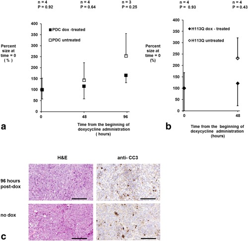
(a, b) Effect of induction of expression of wild‐type (a) and mutant PDC (b) on xenograft growth. The size of the xenografts derived from cells transfected with the tet‐on PDC or tet‐on H113Q constructs are expressed as the percentage of the volume measured at the beginning of doxycycline treatment (0 h). Caliper measurements of two xenograft diameters were made every 48 h from 21 d after implantation, when doxycycline was added to the drinking water as indicated. Euhus’ formula was used to calculate xenograft volume as described in the Methods section. Error bars represent one standard deviation of the mean. Only positive or negative error bars are shown for clarity. (c) Representative formalin‐fixed sections taken from xenografts grown for ∼21–24 d following subcutaneous implantation of cells transfected with the tet‐on PDC‐GFP‐V5/His construct. Where indicated, the mice were given 10 mg/mL doxycycline solution in their drinking water for 96 h prior to xenograft excision. The area of necrotic tissue was assessed via H&E staining, and apoptosis was investigated by probing with an anti‐CC3 antibody. Scale bars = 150 µm.