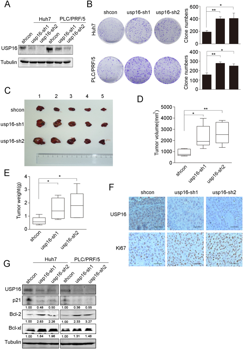
Figure 2. Knockdown of USP16 accelerates the growth of liver tumour cells.
(A) Down-regulation of USP16 by shRNAs (usp16-sh1 and usp16-sh2) in Huh7 and PLC/PRF/5 cells was confirmed by western blot. (B) Images from the colony formation assay are shown (left panel), and the clone numbers for each group were calculated (right panel). (C) Tumours formed by Huh7-shcon or Huh7 shUSP16 cells are shown. (D,E) Tumour volumes (D) and tumour weights (E) were measured. Box-and-whisker plots are shown. Boxes represent the upper and lower quartiles and median; whiskers show the minimum and maximum data points, *p < 0.05, **p < 0.01, determined by t-test. (F) Immunostaining analysis of USP16 and Ki67 in subcutaneous tumours (bar, 100 μm). (G) Huh7 and PLC/PRF/5 with or without knockdown of USP16 were examined by western blotting to test the expression levels of the indicated proteins.

