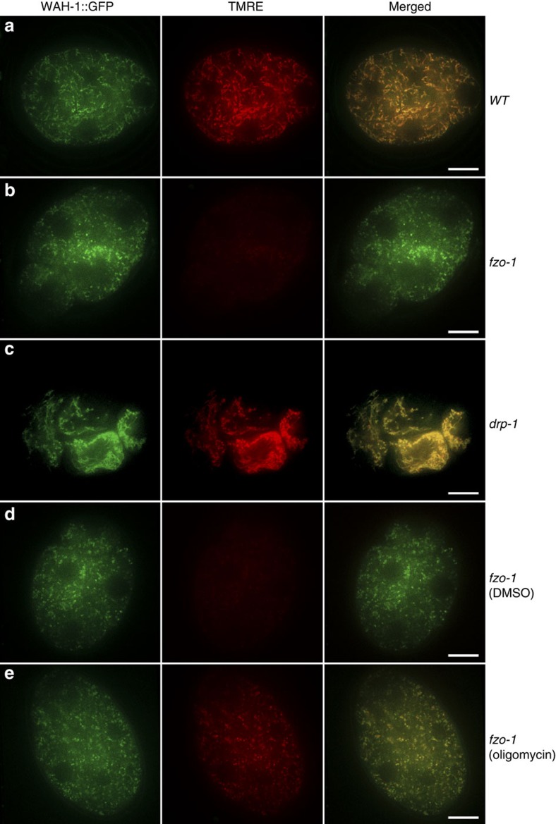
Figure 5. Analyses of mitochondrial membrane potential in maternal mitochondria from different strains.
TMRE staining of 4-cell stage embryos from the following strains are shown: wild type (a), fzo-1(tm1133) (b), drp-1(tm1108) (c), and fzo-1(tm1133) animals treated with DMSO (d) or 150 μg ml−1 oligomycin (e). All strains contain the wah-1::gfp knock-in allele, which was used as an internal mitochondrial marker that labelled both normal and compromised maternal mitochondria. Confocal images of WAH-1::GFP, TMRE and WAH-1::GFP/TMRE merged from embryos dissected from hermaphrodites prestained with TMRE are shown. The exposure time, laser strength and other parameters in each channel are identical for all embryos. The exposure time for the 488 nm laser is 100 ms and for the 561 nm laser is 30 ms. Scale bar represents 10 μm.

