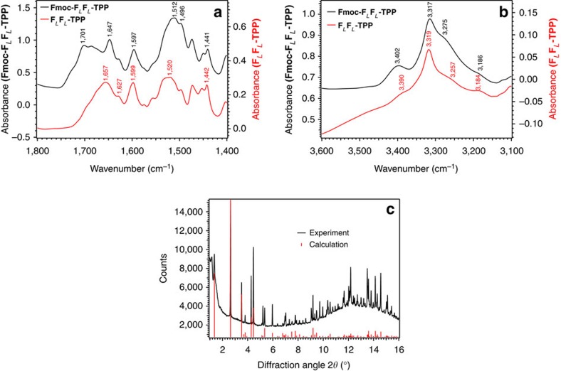
Figure 5. FT-IR spectra and XRD powder diffraction of Fmoc-FLFL-TPP microcrystals.
FT-IR spectra of FLFL-TPP dipeptide KBr pellets in the spectral region of (a) CO, CN, CC and (b) NH stretching vibrations. The black trace is from the Fmoc-protected TPP-FLFL dipeptide while the red trace is from the unprotected FLFL-TPP dipeptide. XRD powder pattern of the same Fmoc-FLFL-TPP microcrystals (c). The black trace is the experimental powder diffraction pattern (λ=1.0000 Å). Red lines are the simulated reflections from the single-crystal data described below. A logarithmic scaling of the measured intensities as a function of 2Θ is presented in Supplementary Fig. 31.

