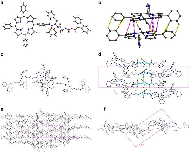
Figure 6. Single-crystal X-ray structure of Fmoc-FLFL-TPP.
(a) Molecule 1. (b) Dimer formed between Molecules 1 and 2 with an enlargement of the two porphyrin moieties (their Fmoc-FLFL units are omitted for clarity) showing C–H···C interactions in yellow and π–π interactions in purple. (c) Another view of this dimer, perpendicular to the porphyrin planes, showing the slipped face-to-face geometry typical to π-stacking in porphyrinic J-aggregates. (d) Intermolecular ‘β-pleated sheet' hydrogen bonding (shown as green dashed lines) between -FLFL- moieties (highlighted in cyan) forming stacks in the b direction. The shifts of the IR frequencies to lower wavenumbers observed in the FT-IR spectra are thus fully accounted for. (e) Interdigitation of porphyrin groups between the β-pleated stacks forming layers parallel to [102], viewed projected onto [102]. (f) as (e) but viewed down the b-axis. Other views are presented in Supplementary Figs 33 and 34 while a larger portion of the crystal lattice is shown in Supplementary Fig. 35.

