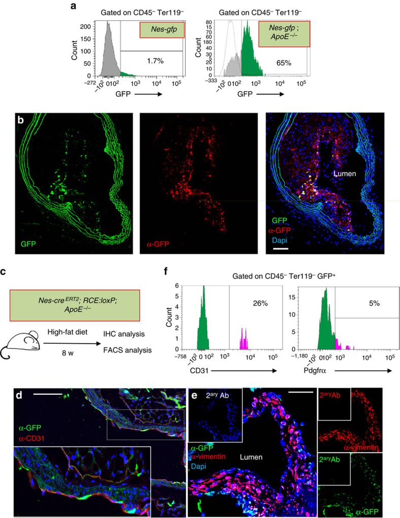
Figure 5. Nestin+ cells participate in the formation of the atheroma plaque.
(a) Flow cytometry of aortic stromal cells showing GFP expression in Nes-gfp mice fed with chow diet (left panel) and in Nes-gfp;ApoE−/− mice fed with HFD for 2 months (right panel). (b) Representative GFP immunofluorescence of a section of the brachiocephalic artery branching out of the aortic arch of Nes-Gfp;ApoE−/− mice fed with HFD for 2 months. Abundant GFP+ cells were found in the atheroma plaque and the adventitial layer. GFP was detected with an anti-GFP antibody (red). Nuclei were counterstained with DAPI (blue). (c) Experimental design of lineage-tracing studies. Nes-creERT2;Rosa26-Gfp;ApoE−/− mice were injected with tamoxifen to trace the progeny of nestin+ cells and were fed with HFD for 2 months. (d) Immunofluorescence of the aortic valves using anti-GFP and anti-CD31 antibodies. Nuclei were counterstained with DAPI (blue). Inset, higher magnification of aortic leaflet. (e) Immunofluorescence of the aortic valves with anti-vimentin antibody. Nuclei were counterstained with DAPI (blue). (f) Flow cytometry histogram showing Pdgfrα and CD31 expression in aortic CD45− Ter119− GFP+ cells. The frequencies of depicted populations are indicated. (b–e) Scale bar, 100 μm.

