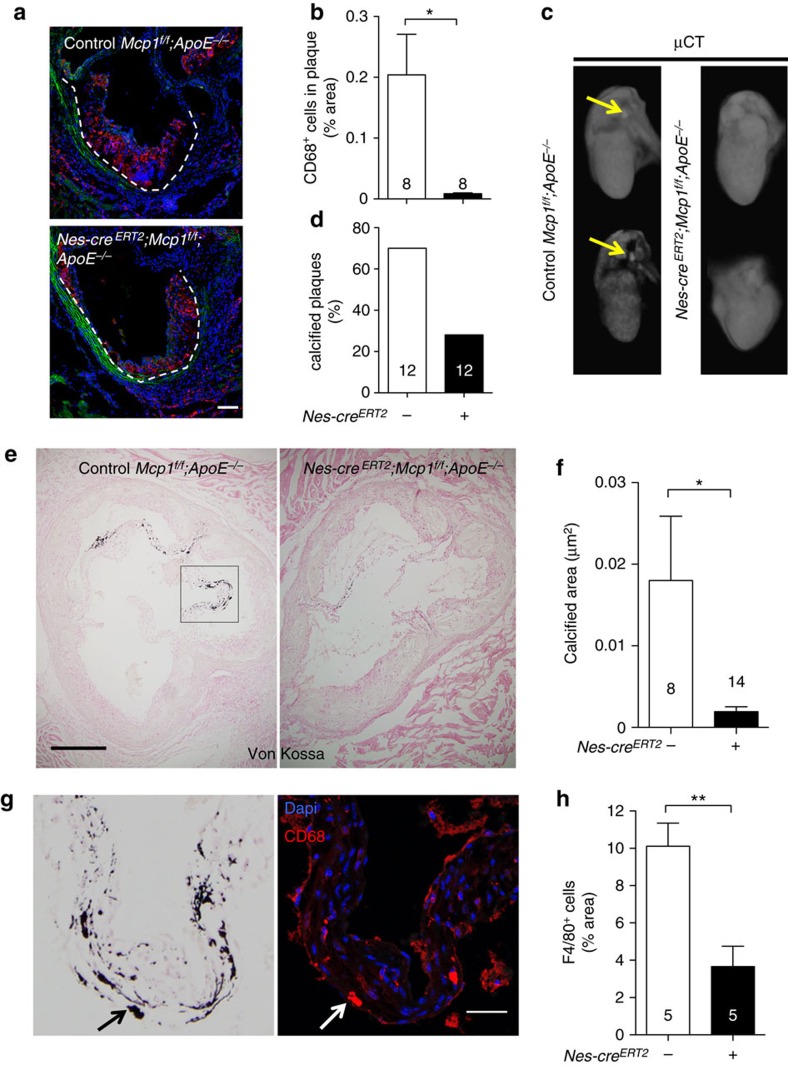
Figure 6. Mcp1 deletion in nestin+ cells reduces vascular calcification.
(a) Immunofluorescence of CD68 (red) to label macrophages infiltrated in the valves of Nes-creERT2;Mcp1f/f;ApoE−/− mice and Mcp1f/f;ApoE−/− controls fed with HFD for 6 weeks. Nuclei were counterstained with DAPI (blue). (b) Quantification of CD68+ cells in the atheroma plaque of these mice (n=8). (c) Representative ex-vivo microtomography (μCT) photographs of the hearts of Nes-creERT2;Mcp1f/f;ApoE−/− mice and Mcp1f/f;ApoE−/− controls. Arrows indicate calcified plaques. (d) Frequency of hearts that showed calcified plaques (n=12). (e) Representative sections of Von Kossa staining of calcium deposits (black) in the aortic valves of these mice. (f) Calcified area in three sections from the aortic valves of these mice (n=8–14). (g) Consecutive aortic valves sections stained with Von Kossa (black) or DAPI (blue) and anti-CD68 antibodies (red). Note the proximity of CD68+ macrophages to the calcified areas (arrow, example). (h) Number of F4/80+ macrophages (not shown) infiltrated in the aortic leaflets (n=5). (b,d,f,h) Data are means±s.e.m.; n and P values are indicated; *P<0.05, **P<0.01, unpaired two-tailed t test. (a, g) Scale bars, 100 μm, (e) 200 μm.

