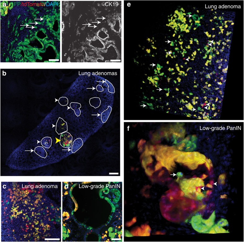
Figure 10. Extra- and intra-tumoral dispersal of early lung and pancreatic tumours.
(a) p53KO/KO (green, GFP+/tdTomato−) high-grade PanIN with extra-tumoral dispersal of CK19-negative cells (arrows) in a 6-week-old Pdx1-Cre-K-MADM-p53 mouse. (b) Stitched confocal image of an entire lung lobe from a 10-week-p.i. K-MADM-p53 mouse reveals multiple low-grade tumours, some harbouring fluorescently labelled cells (arrowheads) and others not (arrows). Most tumours showed dispersed labelling of green and red cells (white arrowheads), whereas some showed clusters of cells (yellow arrowhead). Scale bar, 500 μm. (c) Lung adenoma from a 16-week-p.i. K-MADM-p53 mouse shows dispersed green, red and yellow cells. (d) Low-grade PanIN from a 6-week-old Pdx1-Cre-K-MADM-p53 mouse shows dispersed green, red and yellow cells. (e) Three-dimensional (3D) rendering of multi-photon imaging of a lung adenoma from a 16-week-p.i. K-MADM-p53 mouse showing non-contiguous green (arrows) and red (arrowheads) cells. (f) 3D rendering of multi-photon imaging of a low-grade PanIN from a 6-week-old Pdx1-Cre-K-MADM-p53 mouse showing dispersed green (arrows) and red (arrowheads) cells. Blue, DAPI-stained nuclei. Scale bars, 100 μm (all, unless otherwise noted).

