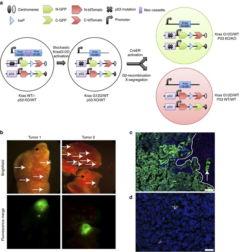
Figure 3. p53 constrains lung tumour progression in the KrasLA2-MADM model.
(a) Schematic of MADM-mediated LOH of p53 in KrasLA2,Rosa26-CreERT2/KrasWT; MADM-p53 mice. Stochastic recombination results in removal of one of two duplicate copies of mutant Kras exon1 (KrasG12D) and an intervening neo cassette permitting expression of mutant Kras expression and tumour initiation25. G2-X MADM recombination, resulting in p53KO/KO (green, GFP+/tdTomato−) and p53WT/WT (red, GFP−/tdTomato+) cells, is initiated through tamoxifen activation of CreERT2, permitting localization of Cre to the nucleus. This diagram was adapted with permission from the original MADM schematic21. (b) Two green tumours (black arrows) were observed on whole-mount analysis of lungs from KrasLA2, Rosa26-CreERT2/KrasWT; MADM-p53 mice (n=8), whereas none were observed in KrasLA2,Rosa26-CreERT2/KrasWT; MADM mice (not harbouring p53 mutation, n=10). White arrows denote tumors without fluorescence labelling. We did not detect any red or yellow tumours in either cohort of mice by whole-mount analysis. Merged fluorescence images of green and red filters are shown. (c) Histologic section of a tumour in b showed green adenocarcinoma cells adjacent to colourless adenoma cells (predominantly to the right of the line). Some green adenocarcinoma cells (arrow) are intercalating in the adenoma area. Blue, DAPI-stained nuclei. Scale bar, 100 μm. (d) KrasLA2,Rosa26-CreERT2/KrasWT; MADM-p53 adenoma harbouring rare yellow cells. Blue, DAPI-stained nuclei. Scale bar, 100 μm.

