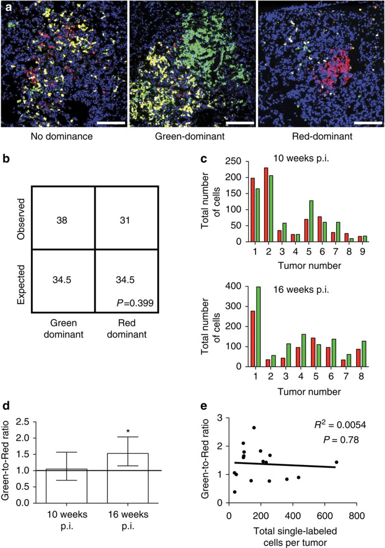
Figure 4. p53 loss does not significantly have an impact on early lung tumorigenesis.
(a) Lung adenomas from K-MADM-p53 mice 16 weeks p.i. showing varying degrees of p53KO/KO (green, GFP+/tdTomato−), p53WT/WT (red, GFP−/tdTomato+) and p53KO/WT (yellow, GFP+/tdTomato+) cell labelling. Representative images of non-dominant, green-dominant and red-dominant tumours are shown. Blue, DAPI-stained nuclei. Scale bars, 200 μm (all). (b) Absolute quantification of observed green-dominant and red-dominant lung adenomas in K-MADM-p53 mice (10–16 weeks p.i., n=5 mice total). Expected numbers are based on 1:1 stoichiometric ratio of green and red cell generation, and stochastic growth thereafter. No statistical difference was observed (P>0.05, χ2-test). A plurality of tumours (51 of 132) did not exhibit colour dominance. (c) Absolute quantification of green and red cells across individual tumours derived from K-MADM-p53 mice evaluated at 10 weeks p.i. (n=9) and 16 weeks p.i. (n=8). (d) Geometric means (±95% confidence intervals) of green-to-red cell ratio in lung tumours (based on data from c, n=9 at 10 weeks p.i. and n=8 at 16 weeks p.i.). Line represents equal green and red cell numbers (ratio=1). The green-to-red cell ratio is mildly but significantly increased in tumours from 16-week-old mice (* denotes 95% confidence interval does not cross unity). (e) No statistically significant correlation between green-to-red cell ratio and total single-labelled (green plus red) cells per tumour was observed (P>0.05, linear regression).

