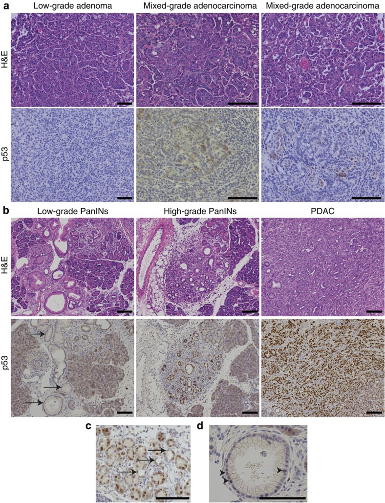
Figure 8. p53 expression in various stages of lung and pancreatic tumour progression.
(a) IHC for p53 in LSL-KrasG12D/KrasWT; p53LSL-R172H/flox low-grade adenomas and mixed-grade adenocarcinomas revealed p53 staining only in high-grade lung tumour cells. (b) IHC for p53 in Pdx1-Cre; LSL-KrasG12D/KrasWT; p53LSL-R172H/WT adult pancreas revealed increased p53 expression in higher-grade pancreatic lesions. Arrows show low-grade PanINs. (c) A subset of acinar-to-ductal metaplasia (ADM) cells expressed p53 (arrows). (d) A subset of low-grade PanIN cells expressed p53 (arrowheads). Scale bars, 100 μm (all).

