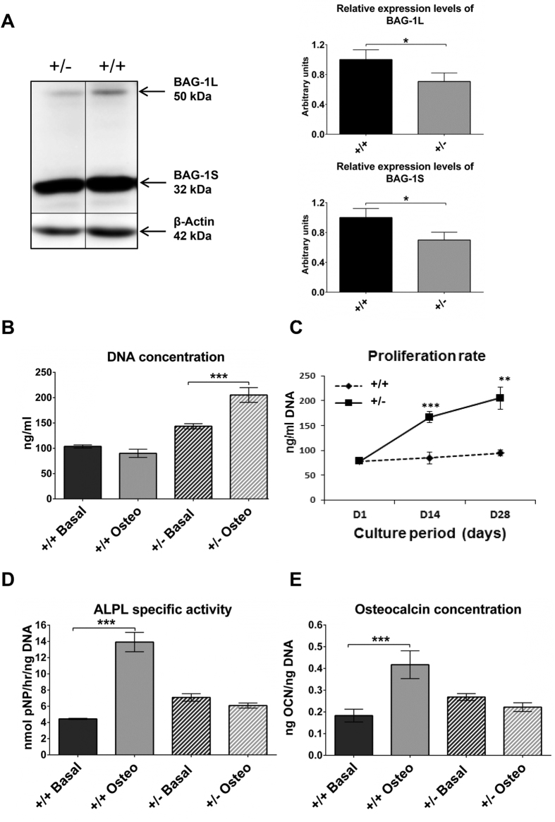
Figure 1. Proliferation and BMP-2-stimulated osteogenic differentiation of BMSCs of 14-week-old Bag-1+/+ and Bag-1+/− female mice.
(A) Representative immunoblots demonstrating bands for BAG-1L, BAG-1S and β-Actin in day-28 cultures of BMSCs of Bag-1+/− and Bag-1+/+ female mice in basal medium. Densitometric quantification of the bands was performed to measure the expression of the BAG-1L and BAG-1S proteins, data was normalised to β-Actin and plotted in the form of bar graphs. (B) DNA concentrations of day-28 cultures of BMSCs of Bag-1+/+ and Bag-1+/− female mice in basal and osteogenic media. (C) Cell proliferation profiles over the course of 28-day osteogenic cultures of BMSCs of Bag-1+/+ and Bag-1+/− female mice. For statistical analyses, DNA concentrations were compared between days 1 and 14 of culture, and days 14 and 28 of culture. (D) ALPL specific activity and (E) osteocalcin concentration were measured in day-28 cultures of BMSCs of Bag-1+/+ and Bag-1+/− female mice in basal and osteogenic media. Results presented as mean ± SD; n = 3 cultures per group; ***P < 0.001, **P < 0.01, *P < 0.05.

